| Search for content and authors |
Monocrystalline character of ZnMgTe shell in the core-shell ZnTe/ZnMgTe nanowires |
| Elzbieta Dynowska 1, Jaroslaw Domagala 1, Przemysław Romanowski 1, Elżbieta Janik 1, Piotr Wojnar 1, Wolfgang Caliebe 2 |
|
1. Polish Academy of Sciences, Institute of Physics, al. Lotników 32/46, Warszawa 02-668, Poland |
| Abstract |
In the case of narrow nanowires (NWs) the surface-to-volume ratio is extremely large and surface states significantly reduce the carrier lifetime and degrade the optoelectronic device performance. Therefore, a surface passivation is of a great importance, especially for ZnTe NWs which oxidize easily. Such passivation can be achieved by forming a shell of a large band gap material around the NW so that the surface states are moved away from the charge carriers confined in the core.
Figure 1. The SEM image of a side-view of the 090110A sample. 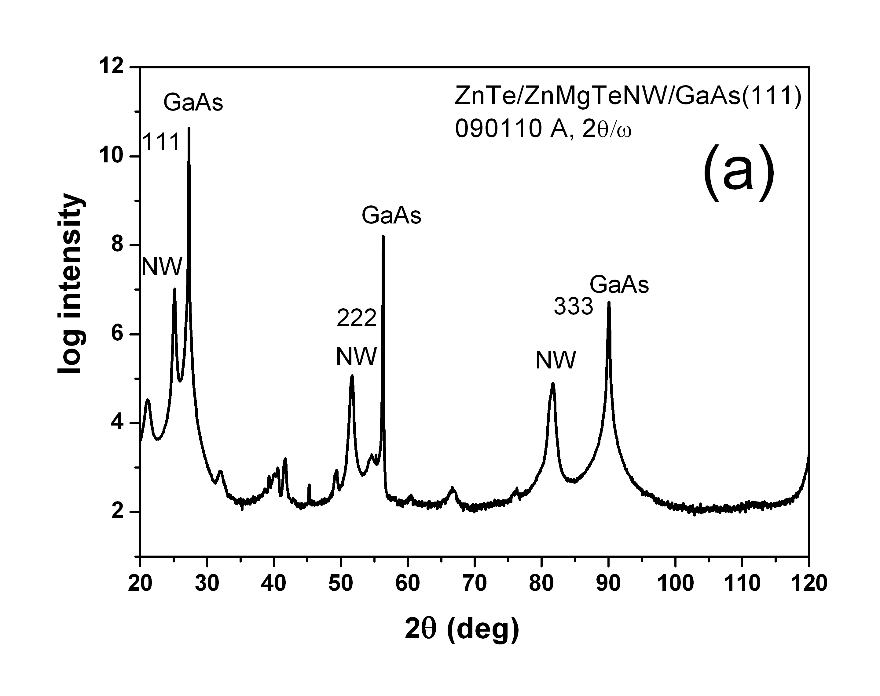 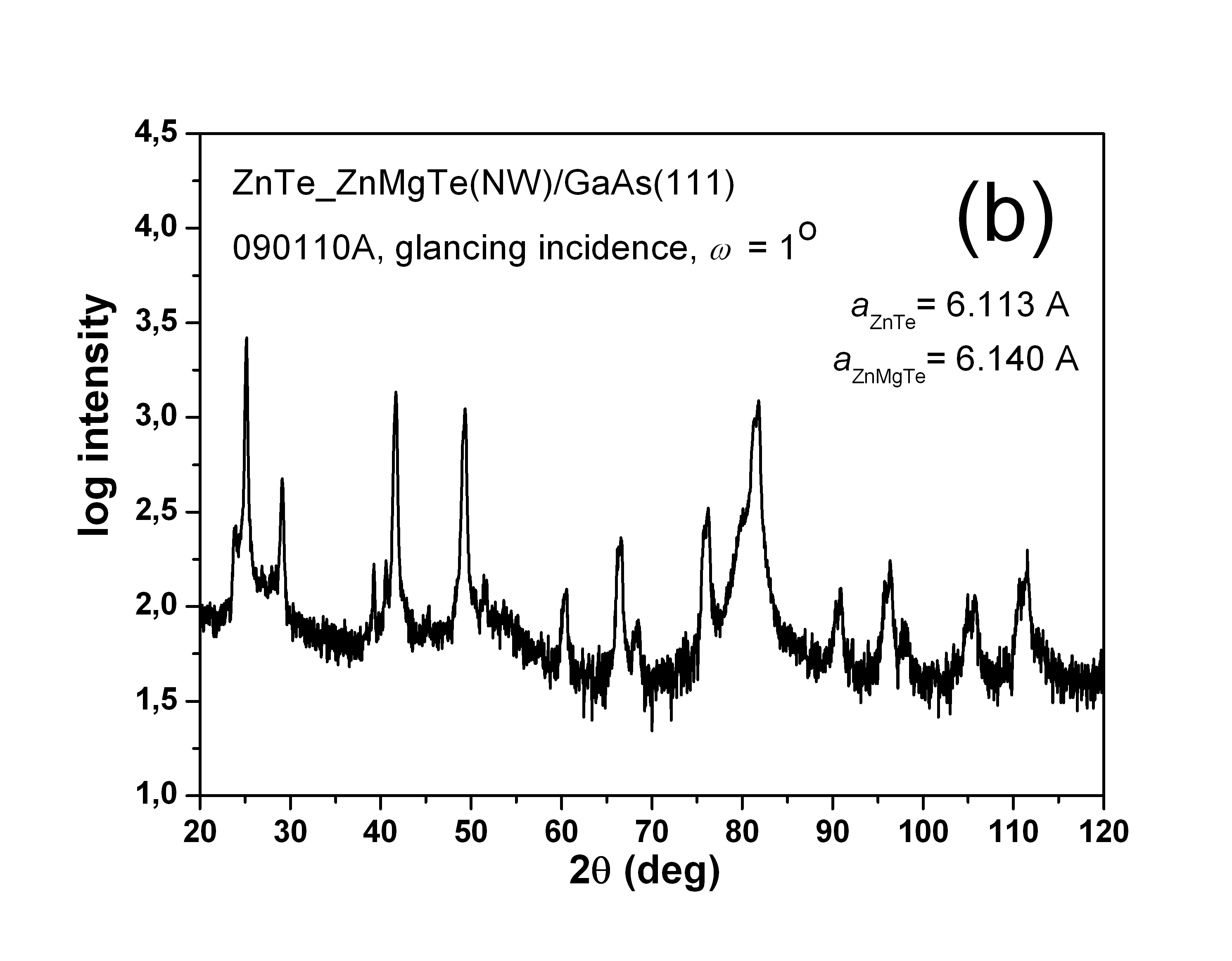
Figure 2. The diffraction patterns of the 090110A sample: (a) symmetrical 2θ/ω scan, (b) 2θ scan in glancing incidence geometry.
In this report we present the results of structural characterization of the core-shell ZnTe/ Zn1-xMgxTe NWs grown by MBE technique on the GaAs(111) substrate. The ZnTe NWs were grown at the temperature of about 470 ºC according to the procedure described in [1]. The Zn1-xMgxTe shells were produced immediately after the growth of the ZnTe cores. In order to produce such shells the substrate temperature was reduced to the value which stops the axial growth of NWs and forces the radial growth of Zn1-xMgxTe shell. The post growth annealing has been applied for some samples. The technological parameters of the investigated samples are listed in the Table 1, and the electron scanning microscopy (SEM) image of a side-view of the 090110A sample is shown in Fig. 1
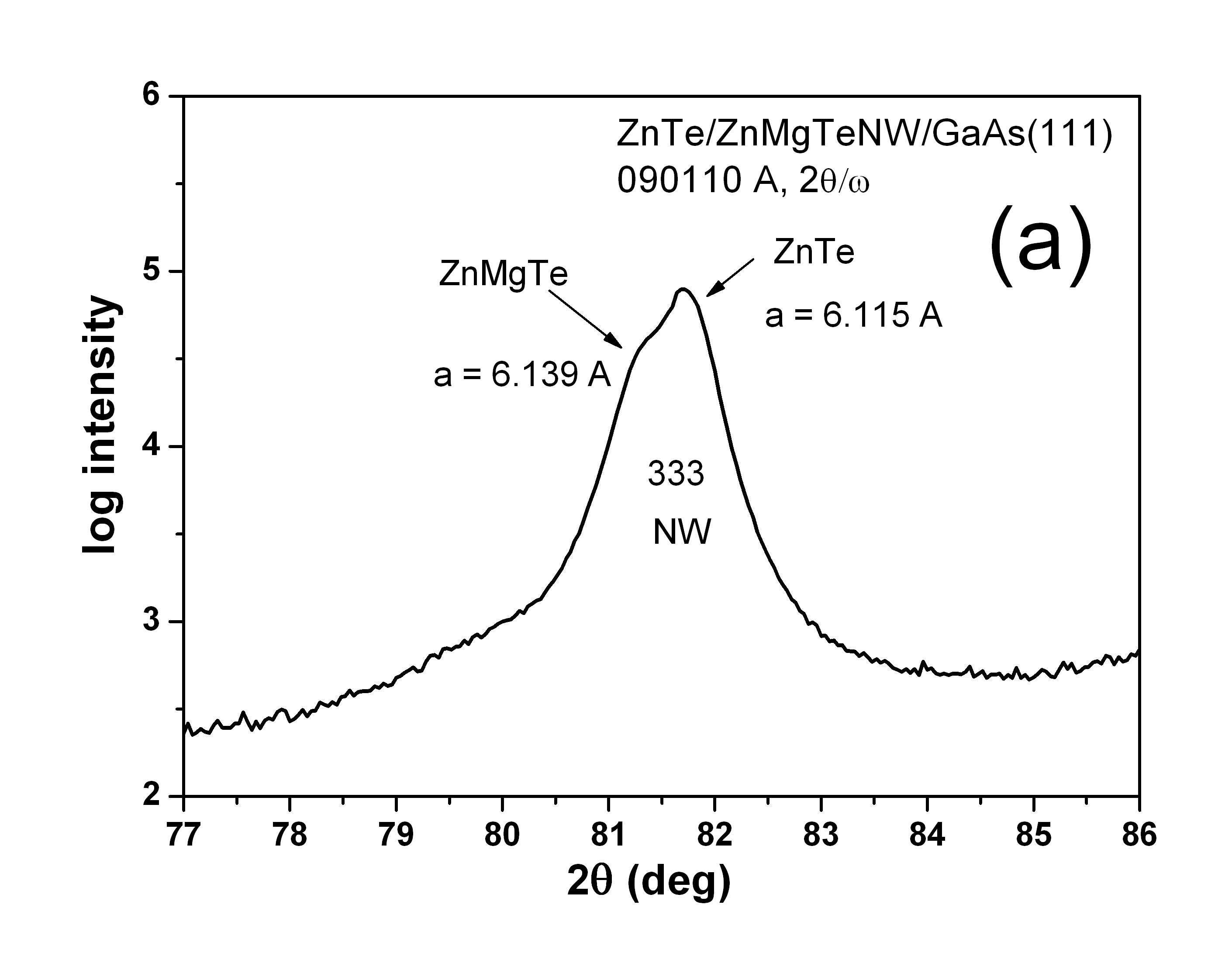
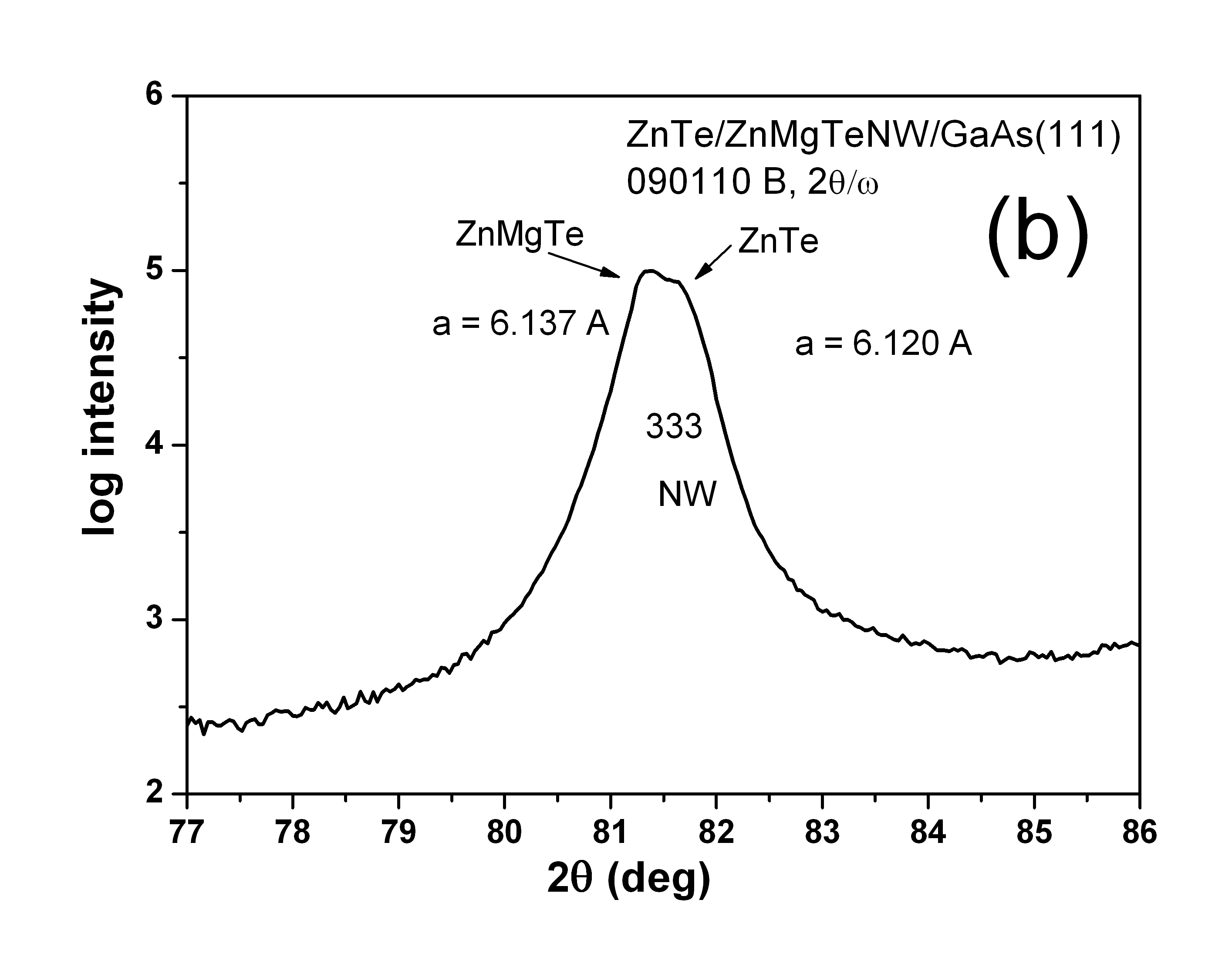
Figure 3. The part of the 2θ-ω diffraction pattern in the vicinity of the 333 NW peaks of the samples 090110A and 090110B with different time of the shell growth: (a) 10 minutes; (b) 15 minutes. The X-ray measurements were performed using synchrotron radiation at the W1 beamline at DESY-HASYLAB. The monochromatic X-ray beam of wavelength λ = 1.54056 Å was used. Two modes of measurement were applied: symmetrical 2θ-ω scan and coplanar 2θ scan in the glancing incidence geometry [2]. The diffraction patterns measured for the sample 090110A in the symmetrical geometry (2θ-ω scan) and in the glancing incidence mode (2θ scan) are shown in Figure 2a,b. They are typical for all studied samples. It was confirmed that the crystallographic orientation of the substrate forces the orientation of NWs – three orders of reflections from (111) lattice planes from GaAs substrate and from NWs are visible in the pattern presented in Figure 2a. The measurement in the glancing incidence geometry (Fig. 2b) shows the relatively thin polycrystalline layer grown on the substrate between NWs. A zoom of the symmetrical pattern in the vicinity of 333 NW peak allows to notice its splitting (see Figs 3 and 4). This splitting indicates the monocrystalline character of the Zn1-xMgxTe shell with the same crystallographic orientation as that of the ZnTe core. 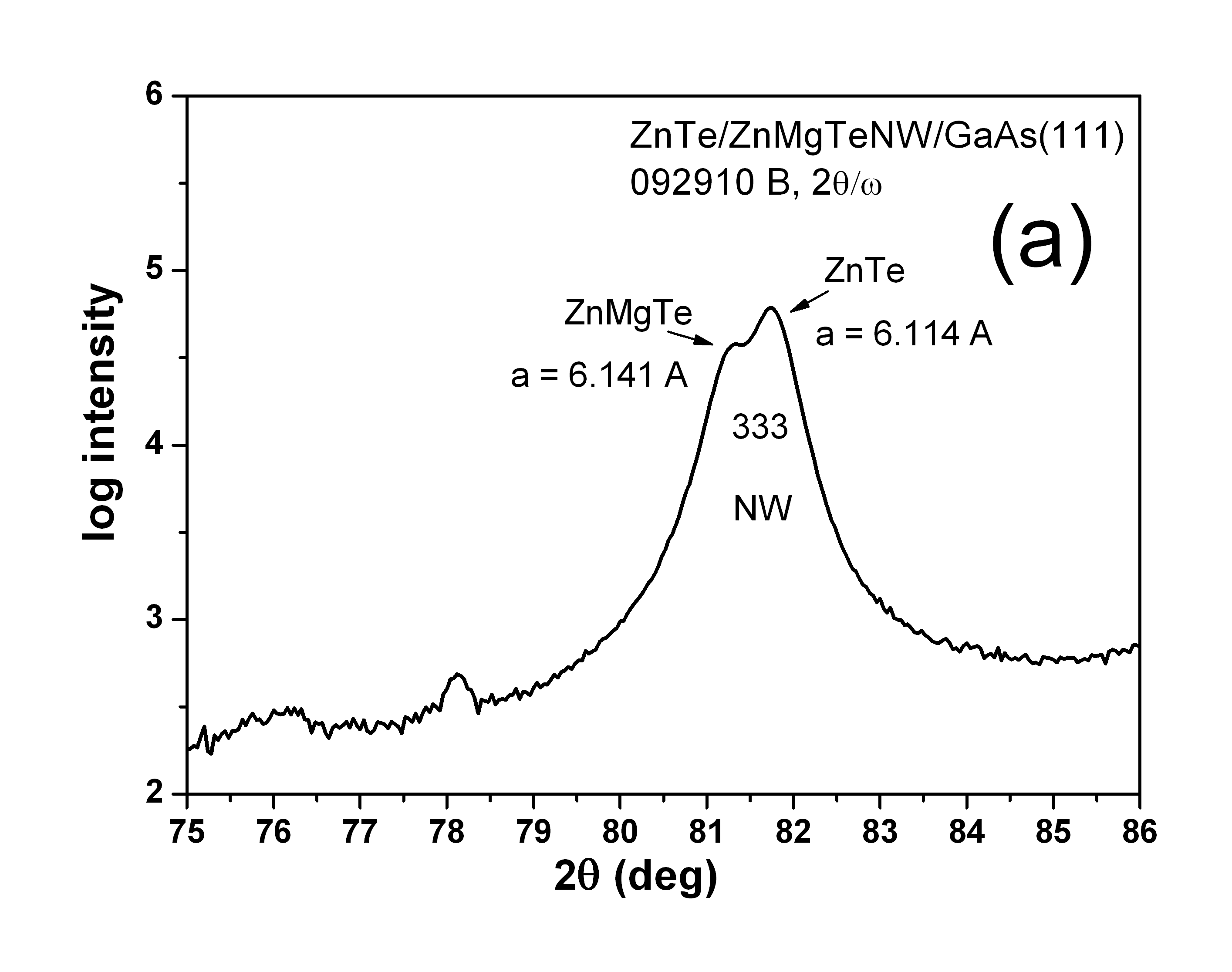 
Figure 4. The part of the 2θ-ω diffraction pattern in the vicinity of the 333 NW peaks: (a) not annealed 092910B sample; (b) sample 092910A annealed at 450 ºC. The second pair of the samples (092910A and 092910B) shows an influence of the post-growth annealing on the NWs structure. In the not annealed sample, 092910B, the splitting of the 333 NW peak is clearly visible (Fig. 4a), while in the annealed sample, 092910A, only small asymmetry on the left side of the peak can be noticed (Fig. 4b), which may be caused by the diffusion of Mg to the core during annealing procedure. From the lattice parameters calculated for Zn1-xMgxTe shell components of the NWs their chemical composition x can be estimated as follows: x ~= 0.11 for the samples 090110A and 090110B and x ~= 0.12 for sample 092910B. In the case of 092910A sample only average value of x can be estimated: xaverage~= 0.075. As it is visible from the Figures 3 and 4, the lattice parameters of the ZnTe cores are larger than that of the uncovered ZnTe NWs [3] – the most probable reason of such behaviour is the tensile strain of ZnTe core caused by Zn1-xMgxTe shell. Acknowledgements: This work was partially supported by the European Community-Research Infrastructure Action under the FP6 "Structuring the European Research Area" Programme (through the Integrated Infrastructure Initiative "Integrating Activity on Synchrotron and Free Electron Laser Science", Contract RII3-CT-2004-506008) and the European Union within European Regional Development Fund, through grant Innovate Economy (POIG.01.01.02-00-008/08). References [1] E. Janik, P. Dłużewski, S. Kret, A. Presz, H. Kirmse, W. Neumann, W. Zaleszczyk, L.T. Baczewski, A. Petroutchik, E. Dynowska, J. Sadowski, W. Caliebe, G. Karczewski, T. Wojtowicz, Nanotechnology 18 (2007) 475606 [2] E. Dynowska, W. Szuszkiewicz, J.Z. Domagała, E. Janik, M. Wiater, G. Karczewski, T. Wojtowicz, W. Caliebe, HASYLAB Annual Reports (2006) 756 [3] E. Dynowska, W. Szuszkiewicz, J.Z. Domagała, E. Janik, A. Presz, T. Wojtowicz, G. Karczewski, W. Caliebe, Radiation Physics and Chemistry 78 (2009) 120 |
| Legal notice |
|
| Related papers |
Presentation: Poster at IX Krajowe Sympozjum Użytkowników Promieniowania Synchrotronowego, by Elzbieta DynowskaSee On-line Journal of IX Krajowe Sympozjum Użytkowników Promieniowania Synchrotronowego Submitted: 2011-06-01 16:20 Revised: 2011-09-19 20:51 |
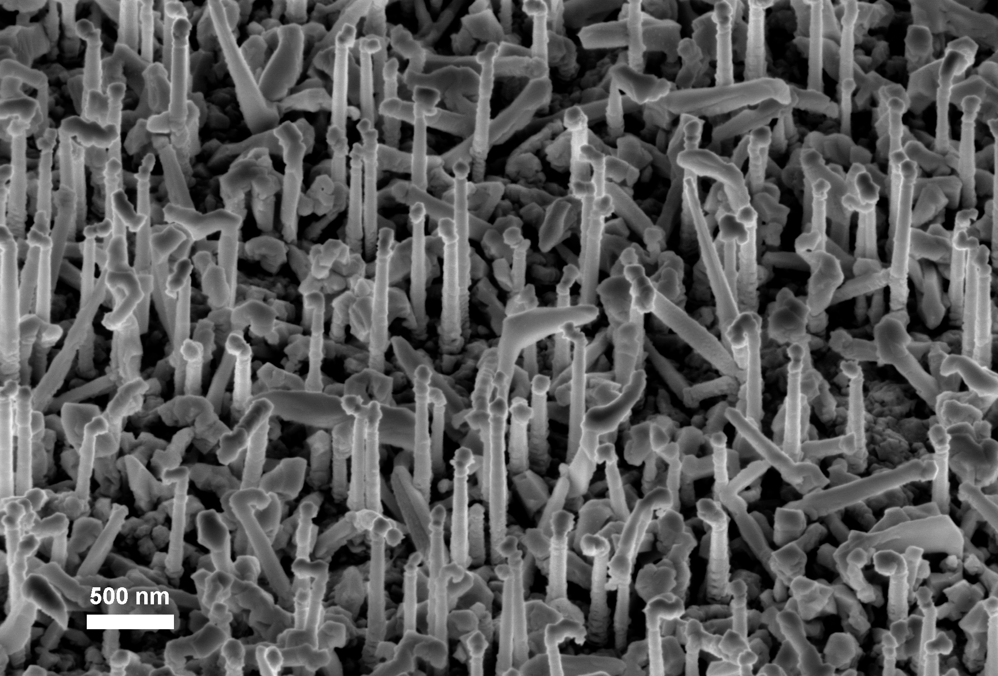
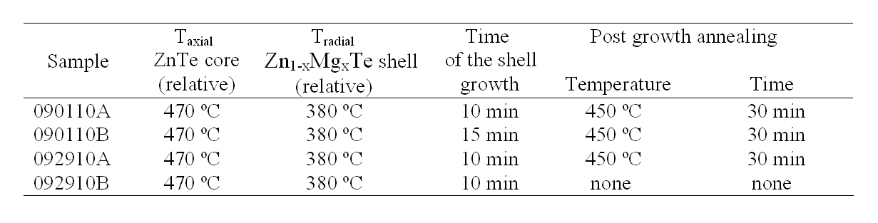 Table 1. The growth and annealing parameters of the core-shell ZnTe/Zn1-xMgxTe nanowires.