| Search for content and authors |
Forming two-dimensional structure of DNA-functionalized Au nanoparticles via lipid diffusion in supported lipid bilayers |
| Takumi Isogai 1, Agnes Piednoir 2, Eri Akada 1, Yuki Akahoshi 1, Ryugo Tero 3, Shunta Harada 1, Toru Ujihara 1, Miho Tagawa 1 |
|
1. Department of Materials Science and Engineering, Nagoya University, Furo-cho, Chikusaku, Nagoya 464-8603, Japan |
| Abstract |
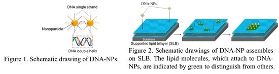
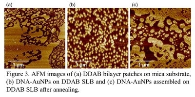
Nanoparticle assemblies have recently attracted considerable interest because of their potential applications in the field of nanoelectronics, nanophotonics and other various nanodevices. To realize novel devices utilizing ‘nano-specific’ phenomena and effects, it is necessary to develop a method for programmable assembly of nanoparticles into one-, two- or three-dimensional arrays with nano-meter precision. In recent years, DNA has attracted much attention as an engineering tool for controlling the assembly of nanoparticles because it offers programmability in the arrangement of nanocomponents through base sequence design. As shown in Figure 1, nanoparticles bind each other via DNA strands, which are attached on nanoparticles, through DNA hybridization: the process of combining two complementary single-stranded DNA molecules and allowing them to form a double-stranded molecule through Watson-Crick base-pairing rules. By this technique, three-dimensional nanoparticle superlattices have already been made using DNA-functionalized nanoparticles (DNA-NPs). To assemble two-dimensional nanoparticle superlattices using DNA-NPs, we figured out to utilize surface adsorption and laterally-confined diffusion of DNA-NPs on a substrate surface. More specifically, to implement laterally-confined diffusion of DNA-NPs, we used supported lipid bilayer (SLB). Figure 2 shows schematic drawings of the process of forming two-dimensional nanoparticle array using lipid lateral diffusion: Firstly adsorption of DNA-NPs to SLB (left), secondly making DNA-NP diffuse via lipid (middle), and finally assembling two-dimensional superlattices of nanoparticles on SLB (right). We prepared two types of DNA-functionalized Au nanoparticles (DNA-AuNPs) as shown in figure 1 and lipid bilayer on mica by vesicle fusion method using Dimethyldioctadecylammonium bromide (DDAB). The solution of DNA-AuNP mixture was deposited on DDAB SLB and temperature of the system was increased in order to promote lipid lateral diffusion. After that, the sample was annealed. Figure 3(a) shows that an AFM image of DDAB SLB on mica. Small bilayer patches of DDAB were observed. Figure 3(b) shows an AFM image of DNA-AuNPs adsorbed on DDAB SLB. Close analysis of section profiles revealed that most of DNA-AuNPs adsorbed selectively on DDAB bilayers. As shown in Figure 3(c), after annealing, the morphology changed dramatically and densely-packed well-ordered two-dimensional arrays of DNA-AuNPs were observed. This result suggests that lipid diffusion was enhanced at high temperature and DNA-AuNPs were transported by high-mobility lipid molecules and then bind each other through DNA hybridization. In this system, the realization of two-dimensionally limited diffusion of DNA-NPs enables two-dimensional assembly of nanoparticles without creating any complex mechanism on nanoparticles to get their own anisotropies.
|
| Legal notice |
|
| Related papers |
Presentation: Oral at 17th International Conference on Crystal Growth and Epitaxy - ICCGE-17, General Session 8, by Takumi IsogaiSee On-line Journal of 17th International Conference on Crystal Growth and Epitaxy - ICCGE-17 Submitted: 2013-04-14 04:56 Revised: 2013-04-15 06:51 |