| Search for content and authors |
The Analysis of Defects in ZnGeP2 Single Crystals from Birefringence Images |
| Alexey Okunev 1, Galina A. Verozubova 2, Chunhui Yang 4, Chong-qiang Zhu 4, Valery Tkal 3, Vladimir Staschenko 1, Inga A. Zhukovskaya 3 |
|
1. Yaroslav the Wise Novgorod State University, Bolshaya St-Petersburgskaya 41, Novgorod the Great 173003, Russian Federation |
| Abstract |
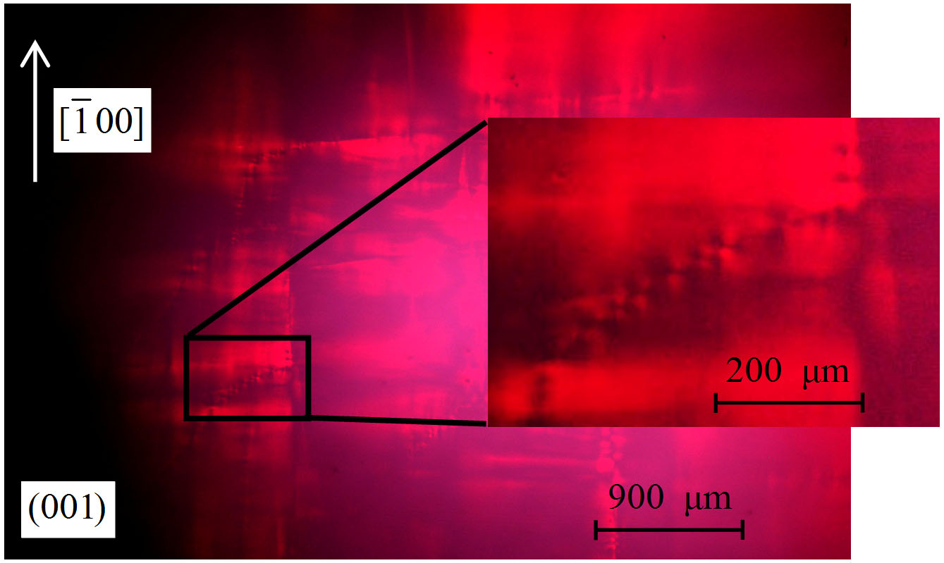
Due to simplicity, exceptional clarity and reliability polarization-optical method (photoelasticity) occupies a special place among all experimental methods of determining of stresses. It is an optical method of stress investigation based on the phenomenon of induced birefringence (piezo-optical effect). It is known that this method allows us to study not only the macroscopic internal stresses in the transversal and longitudinal cuts of single crystal ingots, but also to identify the microscopic stresses associated with individual dislocations [1]. In an optically isotropic crystals dislocations with non-zero edge component of Burgers vector, form a picture of the characteristic stresses, having a form of contrast rosette. Exploring the field of birefringence around the edge or mixed dislocation with line parallel to the axis of observation, we can determine position of the slip plane, the sign and magnitude of the Burgers vector. The method has demonstrated its great possibilities at the study of individual dislocations in silicon, garnet crystals, GaAs and GaP [2]. We studied types of dislocations in SiC single crystals and fine structure of photoelasticity rosettes from them by birefringence method [3, 4]. In some materials (alums, Rochelle salt and corundum) individual dislocations are not resolved and observed images in the photoelasticity method correspond to dislocation clusters or slip lines [2]. The image contrast, and thus the sensitivity of photoelasticity method are determined by many factors: the characteristics of the optical system, the size of the aperture diaphragm of the microscope, the polarizer’s quality, the properties of the photographic material used to fix images, quality of the crystal surface. Despite an advanced theory of characterization of revealed dislocations, visibility and ease of implementation, the method of photoelasticity is relatively rarely applied to the study of structural defects of semiconductors. In this paper, we used the method of polarization-optical analysis to study of structural perfection of the ZnGeP2 single crystals, grown by seeded vertical Bridgman method from melt [5]. Crystals were produced in Institute of Monitoring of Climatic and Ecological Systems SB RAS (Tomsk, Russia) and Single Crystal Laboratory in School of Chemical Engineering, Harbin Institute of Technology (Harbin, PRC). According to our earlier studies carried out by X-Ray topography, these crystals contain dislocations with straight lines [6], and one would expect to find a birefringence patterns from them. Indeed, at study of ZnGeP2 plates, cut along the optical isotropy plane (001), birefringence images have been registered presumably from the slip bands as shown in Fig. 1. Fixed rosettes of photoelasticity from individual dislocations in low-angle boundaries also are inserted in Fig 1. Birefringence image of the entire volume of the sample composed from several single photographs is given in Fig. 2.
Fig. 1. Birefringence image of part of the ZnGeP2 crystal with dislocation rows. 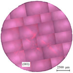
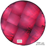
Fig. 2. The whole ZnGeP2 plate, imaged with crossed nicols. After the study by photoelasticity method crystals were thinning by chemical polishing for X-Ray topography. Fig. 3 shows topograph of the same crystal obtained by the method of the X-Ray topography on base of Borrmann effect. It is clear from X-Ray topographs that dislocations are distributed irregularly over the area of the plates, there are almost dislocation-free area of the small size and the areas in which a high density of defects leads to the disappearance of the anomalous transmission of X-Rays due to the strong distortion of the crystal lattice. The average dislocation density is about Nd ~ 5·103 cm-2.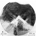
Fig. 3. X-Ray topograph of the same ZnGeP2 plate. Dislocations with curved lines, united in streams like structures and the slip bands are predominated. Comparison of X-Ray topographs and polarization-optical images shows their complete correlation. Birefringence images correspond mainly dislocation clusters in slip bands, the strongest contrast is observed at the boundaries of the slip bands and areas of relatively perfect crystal. As might be expected, the contrast is stronger for more defective crystals, in which large stresses arise. The contrast is also enhanced by increasing of the crystal thickness, since this increases the path length of the ordinary and extraordinary rays, and their path length difference at interference increases too. Large and bright images in the form of rosettes creates by stresses around large inclusions in the sample volume as it is seen from Fig. 4. From a comparison of experimental images follows that similar SiC crystals [4], the method of polarization-optical analysis allows to obtain information, even in the case when the X-Ray topography methods do not work. In areas where the anomalous transmission of X-Rays is absent due to the high dislocation density, Nd> 104 cm2, for example in area in Fig. 1, polarization-optical method allows to fix the defect images, investigate the stress field around them, and determine the characteristics of defects. In particular, the analysis of the form of photoelasticity rosette allows to determine the direction of dislocation line and the direction of the Burgers vector. The sign and magnitude of the Burgers vector are determined by means of optical compensators [1]. Fig. 4. Birefringence image of sample with large inclusions. Studies facilitated by the fact that in the photoelasticity method we can get a continuous series of images of the dislocation at different angles between the dislocation slip plane and the polarization vector of the incident plane-polarized light. In contrast, in the X-Ray topography researcher can get a discrete set of images of the defect on the several "strong" reflections only. Thus, the polarization-optical method was applied to study of defects in ZnGeP2 crystals for first time. It is shown that this method is suitable to investigate the plates, cut perpendicular to the growth axis of the ingots [001]. Images of growth bands and individual dislocations were identified. Comparison of X-Ray and birefringence topographs shows great promise of this method for rapid analysis of the structural perfection of ingots and study of distribution and types of dislocations in them. This work was partially supported by RFBR grant № 12-02-00201. 1. V.I. Nikitenko, Yu.A. Osip'yan, in book: Problems of Modern Crystallography: Collection of Articles in Memory of Academician A. V. Shubnikov (Nauka, Moscow, 1975), pp. 239-261. 2. K. Sangwal Etching of crystals: theory, experiment, and application (Amsterdam, North-Holland, 1987). P. 497. 3. Yu.A. Drozdov, A.O. Okunev, V.A. Tkal, I.L. Shulpina // Zavodskaja Laboratorija. Diagnostika materialov(rus). 2003. Vol. 69, No. 1, pp. 24–29. 4. A.O. Okunev, V.A. Tkal, L.N. Danil'chuk. The study of structure defects of single crystal silicon carbide by direct physical methods (Veliky Novgorod, Yaroslav-the-Wise NovSU, 2006). P. 252. 5. G.A. Verozubova, A.I. Gribenyukov // Crystallography Reports. 2008. Vol. 53, No. 1, pp. 158–163. 6. Okunev A.O., Verozubova G.A., Trukhanov E.M., Dzjuba I.V., Galtier P.R.J. and Said Hassani S.A. // J. Appl. Cryst. 2009. Vol. 42, No. 6, pp. 994–998. |
| Legal notice |
|
| Related papers |
Presentation: Oral at 17th International Conference on Crystal Growth and Epitaxy - ICCGE-17, General Session 5, by Alexey OkunevSee On-line Journal of 17th International Conference on Crystal Growth and Epitaxy - ICCGE-17 Submitted: 2013-03-13 20:35 Revised: 2013-07-28 00:22 |