| Search for content and authors |
Quantitative Criteria of Image Quality Evaluation, Theory and Experiment |
| Valery Tkal 1, Alexey Okunev 2, Anna V. Sharaeva 1, Inga A. Zhukovskaya 1 |
|
1. Saint Petersburg State University of Service and Economics (SUSE), Kavalergardskaja, 7, St-Petersburg 191015, Russian Federation |
| Abstract |
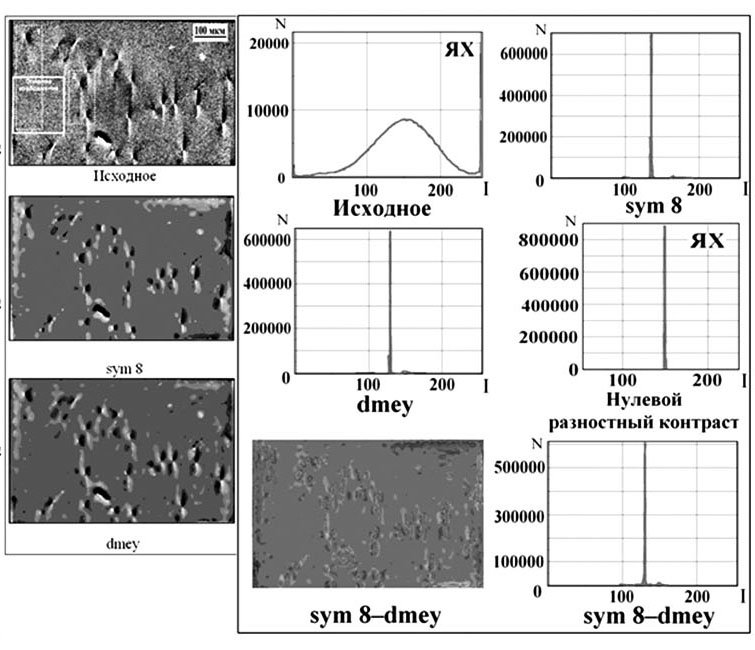
By using different techniques of digital processing and different wavelet bases we can obtain processed images, similar in quality and visually indistinguishable, which actually contain differences due to "thin" features of experimental contrast. Several experts, analyzing the same contrast, may interpret it differently and, therefore, to identify structural defects in different ways. To reduce the influence of the subjective factor, that occurs at visual assessment of contrast, it is necessary to have a simple quantitative criteria for the objective assessment of the quality and efficiency of digital processing, the difference or identity of an analyzed image [1]. To date, existing methodology for quantifying image quality can be divided into two broad categories: subjective and objective techniques. Subjective techniques are used the human visual system to quantify the quality of the images and are based on the median estimate of observers. Objective techniques use mathematical models to simulate the results of a subjective quality assessment, and they based on objectively measurable criteria and metrics. Objective techniques are required repetition of tests to determine the parameters of the processing signal, which reduces the processing speed and complicates these methods. Therefore, it is necessary to find new and simple methods of objective quality assessment. In this contribution, the following objective methods of measuring the quality of images are considered - MSE (Mean Squared Error), PSNR (Peak Signal-to-Noise Ratio), SSIM (Structural Similarity Index). SSIM quality evaluation method is more promising than PSNR and MSE, as the calculation takes into account the brightness, contrast and structural features of the compared images. All of these techniques have a major drawback – they need for time-consuming mathematical calculations, so in this study we used a more simple and easy to implement in practice method, which is based on the construction of brightness characteristics (BC), profiles of intensity (IP) and the difference of contrast (DC) in the program «Image-Pro Plus 6.0» [1, 3]. If one of the two comparable contrast perfectly identical to another, we get a zero DC, and corresponding to it BC and IP have the form of thin vertical and horizontal lines, respectively. If the images have visually indistinguishable minor differences, the DC is not zero, and the maxima of BC are different from ones for zero DC. This approach was tested on the test objects, which were used as a noisy and not noisy theoretical picture of structural defects further subjected to digital processing with different wavelets. Obtained results for the test objects are fully coincides with the results of digital processing of the experimental contrast. For simulation of noise factors – emulsion graininess and background inhomogeneity the expression s2 = s1 + ((Random (10) / 10) - (5/10)) was use. Example of digital processing by different wavelets of noised after simulation test contrast of screw dislocation is shown on Fig. 1. The Fig. 1. shows the results of a similar processing of the experimental topograph of 6H-SiC crystal. The differences are revealed only when comparing of DC and maximum values of BC. Fig. 1. Results of digital processing by dmey and sym 8 wavelets of test image, based on the construction of the DC, BC and IP. The arrow shows the direction of the IP construction. Fig. 2. Results of digital processing by dmey and sym 8 wavelets of experimental topograph. Maxima of BC in the case of processing of test (noised) and experimental contrasts were higher and closer to value for the initial contrast when choosing wavelet basis sym 8 than for the wavelet basis dmey, and difference of contrast "sym 8 - dmey" is not zero. Consequently, digital processing using wavelet sym 8 gives better results than using wavelet dmey. This method was successfully tested on the polarization-optical images with a strong background inhomogeneity, for comparing the effectiveness of different digital techniques and also for images presented in the 8-, 16- and 32-bit format, etc. Thus, it is shown that the use of quantitative evaluation the quality of experimental contrast formed single crystal structure defects, can significantly reduce the subjective factor in its visual estimation and to avoid errors in the identification and localization of defects in crystal volume. Acknowledgments. This work was performed in joint with Ioffe Physical Technical Institute RAS Laboratories "Computer technology in the diffraction diagnostics of materials" of SPbSUSE (Novgorod branch) and "X-ray topography methods at research of materials for electronic engineering" of NovSU with the support of RFBR grant № 12-02-00201. 1. V. Tkal, A. Okunev, I. Zhukovskaya, The Brightness and Frequency Analysis of Images of Structure Defects (LAP LAMBERT Academic Publishing, Saarbrücken, 2012), p. 392. 2. V.A. Tkal', I.V. Dzyuba, L.N. Danil'chuk. Computer simulation of the theoretical contrast of various types of structure defects with noise factors. Journal of surface investigation: X-ray, synchrotron and neutron techniques. 2009. V3. No 5. P. 797-803. 3. V.A. Tkal, Quantitative criteria for efficiency evaluating of digital processing of single crystal structure defect images. Proceedings of "X-ray optics – 2010", Chernogolovka, IMT RAS. 2010. P. 37-39. |
| Legal notice |
|
| Related papers |
Presentation: Poster at 17th International Conference on Crystal Growth and Epitaxy - ICCGE-17, General Session 5, by Alexey OkunevSee On-line Journal of 17th International Conference on Crystal Growth and Epitaxy - ICCGE-17 Submitted: 2013-03-13 20:49 Revised: 2013-04-09 21:25 |