| Search for content and authors |
Epitaxial growth of molecules and salts on muscovite mica |
| Wester De Poel 1, G. S. Pintea 2, S. Bozelie 1, J. Münninghoff 1, D. Lensen 1, J. Drnec 2, F. Carla 2, R. Felici 2, P. Mulder 1, J.A.A.W. Elemans 1, W.J.P. van Enckevort 1, A.E. Rowan 1, E. Vlieg 1 |
|
1. Radboud University Nijmegen, Toernooiveld 1, Nijmegen 6525ED, Netherlands |
| Abstract |
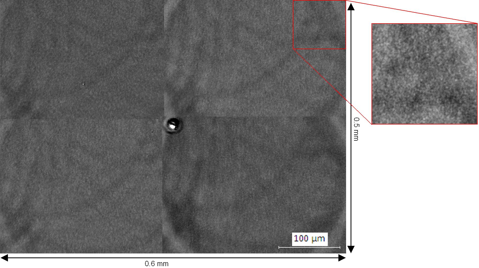
| Muscovite mica is a very commonly used material for scientific study because of its atomically flat surface, which is easily prepared by cleavage. To see how flat muscovite mica really is we examined the surface of this crystal using atomic force microscopy (AFM), phase contrast microscopy and surface X-ray diffraction (SXRD). AFM revealed only one step in over 2500 unique measurements of 2.5 by 2.5 micrometer surface areas. This indicates that the occurrence of steps is very rare. Phase contrast optical microscopy was used to investigate larger surface areas (figure 1)and showed that areas the size of 1mm2 exist that are atomically flat. To investigate even larger surface areas SXRD was used, employing an X-ray beam footprint of 3 by 3 millimetres. Steps can be detected because of the difference in orientation between two neighbouring potassium layers separated by a 1 nm step, as a result from the glide plane in the crystal structure. Using the symmetric (11) and (1-1) crystal truncation rods, SXRD revealed that areas of 5 by 5 millimetres in size exist that are atomically flat. When this flatness is compared to the surface area of the USA, there would only one step dividing the country of two meters in height.
Figure 1 Phase contrast microscopy image of muscovite mica, showing a step-free area of 0.6 by 0.5 millimetres. The rings are an artefact from the microscope, the lines in the middle, both horizontal and vertical, are a result from merging four images and the spot at the centre is a dust particle. The rare occurrence of steps on the muscovite surface means that step-induced nucleation is negligible to epitaxial nucleation on the flat areas of the substrate. Hence, growth in all directions is equally likely and epitaxy is easier to detect. For this reason muscovite mica is suited to grow epitaxial salt crystals. A large range of salts was investigated and analyzed with AFM and optical microscopy. The crystallographic plane with which the salt coordinates to the muscovite was established using X-ray diffraction and from these measurements a relationship between lattice mismatch and the occurrence of epitaxy could be determined.
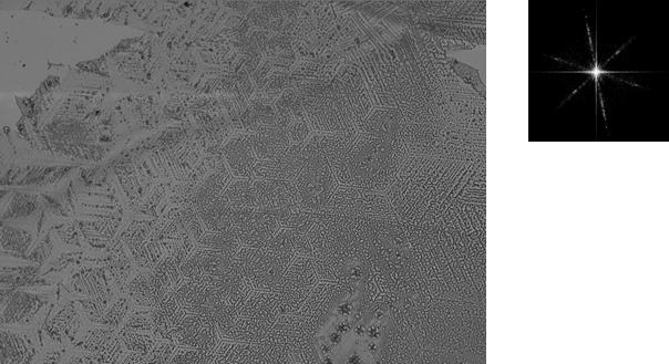
Figure 2 Optical microscopy image of epitaxially grown silver nitrate on muscovite mica (left) and Fourier transform showing the six fold symmetry of the deposit. Finally, molecular monolayers and epitaxial layers were produced on muscovite mica. Crown-ethers are known for their ability to specifically bind to certain metal ions. Muscovite mica has potassium ions at its surface, while dibenzo-18-crown-6 specifically binds to potassium. This molecule should therefore form monolayers, which was indeed observed with AFM. The charge on the surface of muscovite can also be used to produce molecular layers, by binding molecules which have a dipole moment or contain acidic groups. Molecular (epitaxial) monolayers have been produced in this way with porphyrins and benzene-derived molecules.
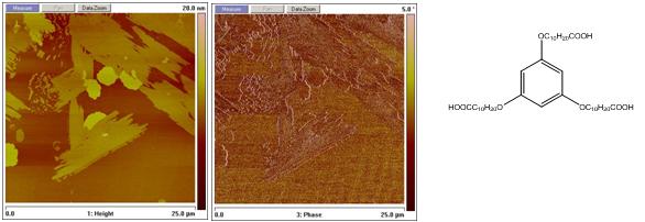
Figure 3 AFM height (left) and phase (middle) images of epitaxial layers of a benzene-derivative (right). The final aim of this project is to use the epitaxial molecular monolayers as a template for protein crystallization. |
| Legal notice |
|
Presentation: Oral at 17th International Conference on Crystal Growth and Epitaxy - ICCGE-17, General Session 10, by Wester De PoelSee On-line Journal of 17th International Conference on Crystal Growth and Epitaxy - ICCGE-17 Submitted: 2013-03-20 14:27 Revised: 2013-07-23 17:00 |