| Search for content and authors |
Characterization of local rocking curves of protein crystals by X-ray digital topography with CCD camera |
| Takeharu Kishi 1, Daiki Fujii 1, Shiro Tsukashima 1, Kei Wako 2, Kenichi Kojima 2, Masaru Tachibana 1 |
|
1. Yokohama City University, Yokohama, Japan |
| Abstract |
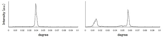
X-ray topography is one of the most powerful methods to characterize the imperfection or crystal defects in protein crystals. Recent development of high resolution CCD camera has led to X-ray digital topography which can acquire a lot of topographs as digital data in short time, depending on the rotation angle of the crystal sample around the Bragg angle for an interest reflection [1,2]. From the analysis of the digital data, we can plot X-ray intensities as a function of the sample rotation angle for any local areas in the topographs, called local rocking curves. Such analysis can lead to more detailed evaluation of the imperfection in protein crystals. In this paper, we report the characterization of local rocking curves of hen egg-white lysozyme (HEWL) crystals with monoclinic structure. Monoclinic HEWL crystals were grown by a batch method and a liquid-liquid interfacial precipitation method. X-ray digital topography with CCD camera was carried out using synchrotron radiation in BL15B1 or 15C at PF in KEK. The wavelength of the incident beam was 1.2Å. The successive digital topographs were recorded in the interval of 0.00053 or 0.001 degree for the analysis of rocking curves. Fig. 1 shows typical local rocking curves for monoclinic HEWL crystals. One is single peak and the other is double peak. In general, the full width of the half maximum of the single peak can be related to the mosaic structure. The double peak might be associated with larger sectors including mosaic structures. The difference between peak positions would correspond to misorientation between sectors with mosaic spreads. The characterizations of local rocking curves will be discussed and compared with other polymorphism such as tetragonal and orthorhombic structures.
Fig. 1 A single peak (left figure) and a local rocking curve which peak was divided (right figure) in a monoclinic lysozyme crystal. Reference Corresponding author: Masaru Tachibana, Yokohama City University, 22-2 Seto, Kanazawa-ku, Yokohama 2360027, Japan, Tel &Fax: +81-45-787-2360, E-mail: [email protected] |
| Legal notice |
|
| Related papers |
Presentation: Poster at 17th International Conference on Crystal Growth and Epitaxy - ICCGE-17, General Session 3, by Masaru TachibanaSee On-line Journal of 17th International Conference on Crystal Growth and Epitaxy - ICCGE-17 Submitted: 2013-04-14 11:38 Revised: 2013-07-17 13:27 |