| Search for content and authors |
Stabilities of Crystal Faces of Calcite Compared by AFM Observation of Facet Formation Processes during Dissolution in Dilute Aqueous Acetic Acid |
| Hitoshi Shindo 1, Yohei Ohtsu 1, Ryohei Yamamura 1, Tamon Kose 1, Kaori Niki 2 |
|
1. Chuo university, 1-13-27 Kasuga, Bunkyo-ku, Tokyo 112-8551, Japan |
| Abstract |
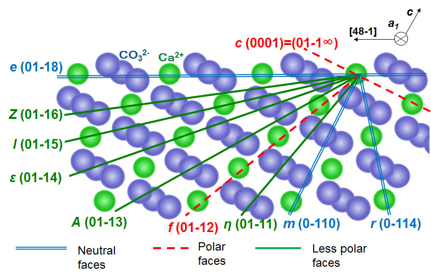
Calcite(CaCO3) crystals are obtained in various forms in nature depending upon their origins[1]. The mineral, accordingly, is a good target in studying relationship between crystal morphology and the growth conditions. A series of crystal faces around the a1-axis are shown in Figure 1. Depending on the inclination from the basal plane, electrically neutral e-, m- and r-faces, polar c- and f-faces, and other less polar faces are recognized. Many of them appear in natural crystal forms. Electrical polarity greatly affects stabilities of the crystal faces in polar solvents like water.
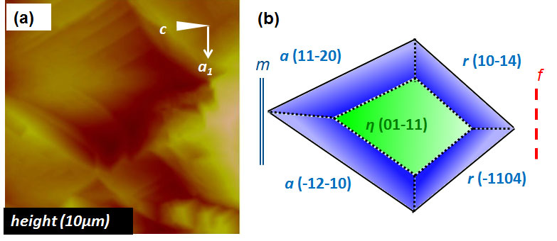
Figure 1. Structure of calcite crystal viewed along the a1-axis. Hexagonal unit cell with the lattice constants of a =499.0 and c =1706.1 pm was chosen. To study the relationship between crystal morphology and the growth conditions, we need to evaluate thermodynamic stabilities of various faces of crystals. We can experimentally compare relative stabilities of crystal faces, by observing, with atomic force microscopy(AFM), the formation processes of polyhedral micro-etch pits at various starting faces. Since diffusion is limited within tiny etch pits, especially for less soluble crystals, near equilibrium is obtained. By starting with a less stable crystal face, we can observe development of sidewalls formed by more stable facets. If we start with locally the most stable surface, atom-flat terraces are often obtained. The method was previously employed in studying crystal morphologies of aragonite-type carbonates[2-4], anhydrite(CaSO4)[5], and NaCl[6]. Calcite crystal faces in desired orientations were prepared by cutting natural calcite crystals buried in resin with Leica SP1600 microtome having a doughnut-shaped diamond saw. Fine-polished crystal plates in m(10-10) orientation were purchased from Furu-Uchi Chemicals. All the crystal faces shown in Figure 1 were prepared. The crystal plates were soaked in dilute aqueous acetic acid or water in a flow-cell for AFM. When required, ethanol was added to the solvent to decrease polarity. The dissolution processes were observed with NanoScope III AFM of Digital Instruments in contact mode. Directions of ledges formed and dihedral angles between the sidewalls and the starting surface tell us the Miller indices of the facets stabilized in the solution. Figure 2(a) shows an AFM image observed when η(01-11) face was dissolved in 1 mM aqueous acetic acid for 3 hours. Development of flat terraces shows us that the starting face is fairly stable. As shown in the sketch in (b), electrically neutral and stable r-faces form two sidewalls on the right-hand side. On the left-hand side, neutral a-facets give macro-steps and sidewalls. On the other hand, neutral but rugged m(01-10) face and polar f(01-12) facets do not appear in (a). We can tell that η-, r- and a-facets are more stable than m- and f-facets. By changing the starting faces, relative stabilities of calcite faces in aqueous acetic acid were determined as r > e > η, ε, a > m > c, f. Polar c- and f-faces are not stable in the solution.
Figure 2. (a):AFM image observed during dissolution of η(01-11) face of calcite in 1mM aqueous acetic acid and (b): a sketch of an etch pit formed. More stable η-, r- and a-facets make the terraces and sidewalls, while less stable m- and f-facets do not appear in (a). When ethanol was added to the aqueous acetic acid, however, polar c- and f-facets become much more stable. Adsorption of surface active ethanol molecules most probably stabilizes the polar faces. The polar faces often appear in natural crystal forms. The changes in the relative stabilities of crystal faces give us clues to the relationship between crystal forms and growth conditions. Acknowlegement This work was supported by JSPS KAKENHI 24510144, 20510097, 20225002, and The Institute of Science and Engineering, Chuo University. References [1] E. S. Dana, The System of Mineralogy –Descriptive Mineralogy, Wiley, New York, 6th ed. 1900, p.262. [2] M. Kwak and H. Shindo, J. Cryst. Growth, 275 (2005) e1655-1659. [3] H. Shindo and M. Kwak, Phys. Chem. Chem. Phys., 7 (2005) 691-696. [4] Y. Shirota, K. Niki and H. Shindo, J. Cryst. Growth, 324 (2011) 190-195. [5] H. Shindo, T. Igarashi, W. Karino, A. Seo, M. Yamanobe-Hada and M. Haga, J. Cryst. Growth, 312 (2010) 573-579. [6] W. Karino, H. Koda, K. Nakamura and H. Shindo, J. Cryst. Growth, 310 (2008) 676-681. |
| Legal notice |
|
| Related papers |
Presentation: Oral at 17th International Conference on Crystal Growth and Epitaxy - ICCGE-17, General Session 9, by Hitoshi ShindoSee On-line Journal of 17th International Conference on Crystal Growth and Epitaxy - ICCGE-17 Submitted: 2013-03-27 06:22 Revised: 2013-07-17 18:41 |