| Search for content and authors |
Structural characterization of a coarse-grained transparent silicon carbide powder by a combination of powder diffraction techniques |
| Burkhard Peplinski 1, Andy Fitch 2, Alexander Evans 2, Richard M. Ibberson 3, Daniel M. Többens 4,5, Lachlan M. Cranswick 6, Ilona Dörfel 1, Franziska Emmerling 1, Ralf Matschat 1 |
|
1. Federal Institute for Materials Research and Testing (BAM), Richard-Willstätter-Str. 11, Berlin D-12489, Germany |
| Abstract |
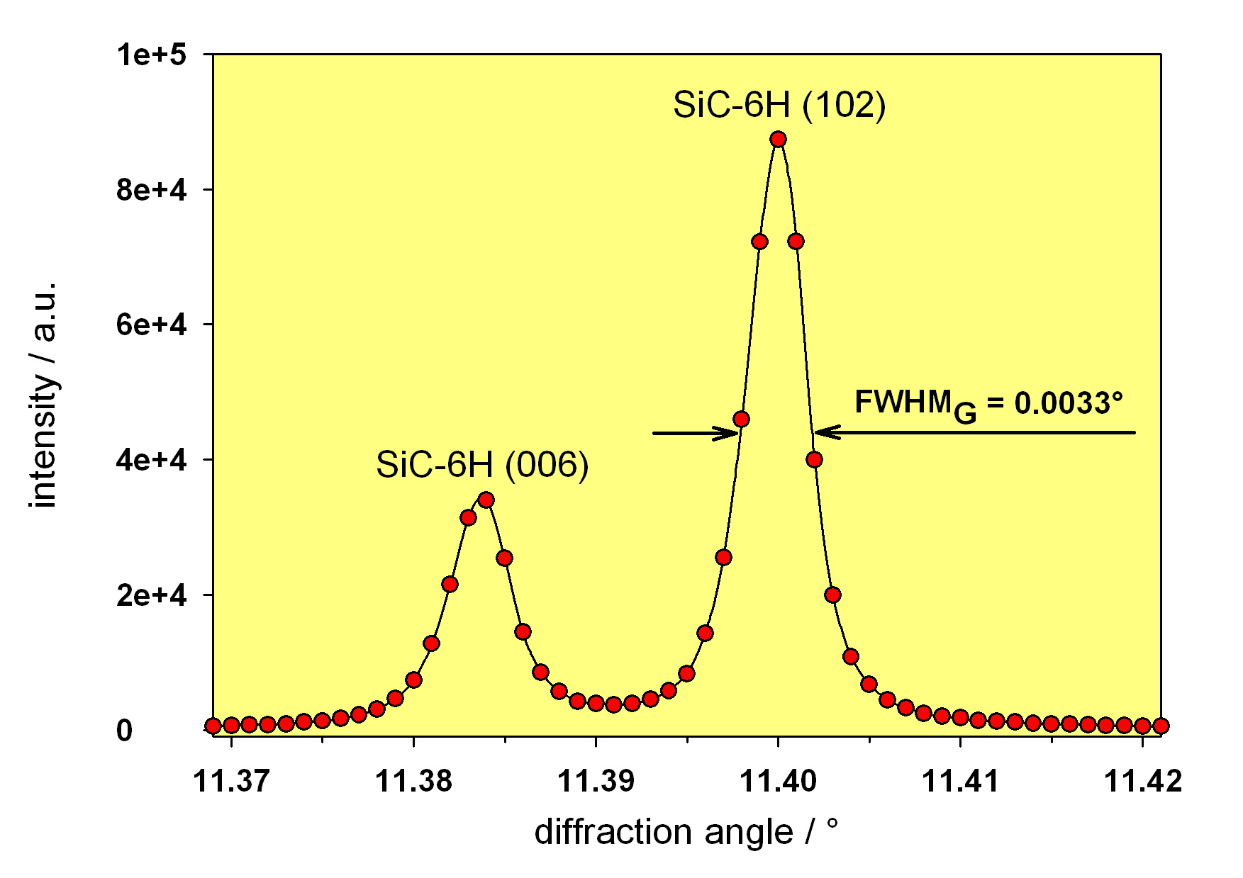
Silicon carbide powders having extremely low levels of chemical impurities (a few μg per g or even less), high perfection of the crystalline lattice and a grain size of up to a hundred μm show exceptional chemical resistance (insolubility in any kind of acid), high hardness as well as unique electronic and optical properties. Along with the lack of reliable analytical techniques for the quantitative determination of chemical trace impurities in such materials, their structural characterization is very challenging, too. One of the main problems is the considerable discrepancy between the actual grain size of the coarse-grained SiC powder samples and the ideal size for powder diffraction measurements. Any grinding bears a high risk of changing/destroying the original real structure characteristics of the sample, e.g. the degree of stacking disorder and the polytype composition. Thus, grinding can significantly distort the outcome of the structural investigation and, therefore, should be avoided. In an attempt to characterize the structural properties of such a material as fully as possible powder diffraction data of a large number of samples were collected applying conventional X-ray diffractometry (Bragg-Brentano geometry), constant-wavelength neutron diffractometry, time-of-flight neutron diffractometry and finally synchrotron radiation (SR) diffractometry at a dedicated beamline. All neutron and SR diffraction measurements were carried out on capillary specimens. Data evaluation was carried out with the program packages TOPAS, GSAS and FULLPROF. The present investigation demonstrates how the specific strengths of each of these four non-destructive powder diffraction techniques complete each other very nicely. Their results go together with the outcome of a TEM investigation for which individual grains of the powder sample were thinned into 100 nm thick slices by the focused ion beam (FIB) sample preparation technique.
|
| Legal notice |
|
| Related papers |
Presentation: Poster at 11th European Powder Diffraction Conference, Poster session, by Burkhard PeplinskiSee On-line Journal of 11th European Powder Diffraction Conference Submitted: 2008-04-30 10:27 Revised: 2009-06-07 00:48 |